
Latest Blog
Zinc is a vital trace element. The body cannot produce the nutrient itself and can only store limited amounts of it. Therefore, it is important to meet your daily requirement
All Blog Posts
Zinc is a vital trace element. The body cannot produce the nutrient itself and can only store limited amounts of it. Therefore, it is important to meet your daily requirement
If the body lacks vitamin D3, this is noticeable with numerous deficiency symptoms. Possible symptoms of a persistent vitamin D3 deficiency include increased susceptibility to infections and muscle weakness.
Vitamins D3 and K2 complement each other. Especially if you take a high-dose vitamin D3 supplement, a combination with vitamin K2 is recommended to minimize the risk of calcium deposits
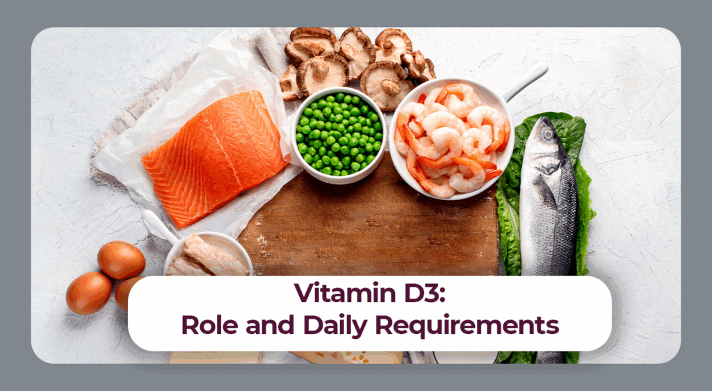
Vitamin D or vitamin D3 has many important functions. Among other things, it is indispensable for bone and tooth mineralization. The body needs sunlight to be able to produce the

Vitamin B2 or riboflavin is contained in both animal and plant foods. Those who eat a balanced diet therefore usually consume enough for their individual needs.
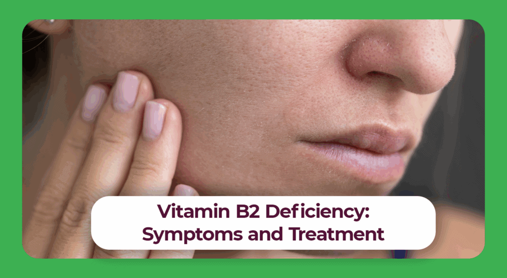
In most cases, a vitamin B2 deficiency is triggered by a health problem. Vitamin B2 preparations are used to treat the deficiency symptoms associated with it.